

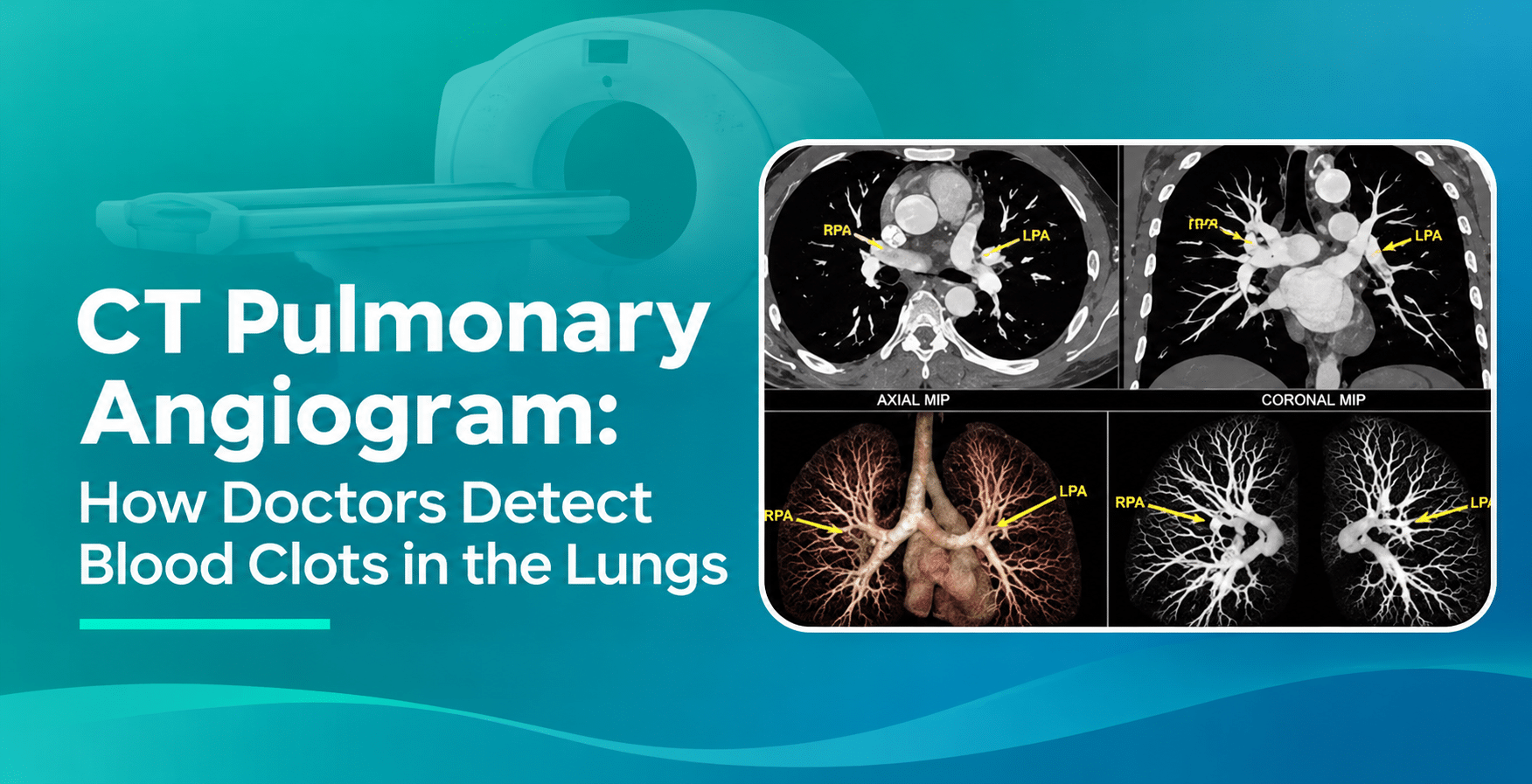
CT Pulmonary Angiogram: How Doctors Detect Blood Clots in the Lungs
Need a CT Angiogram in Dubai ? Contact Us Now
A CT pulmonary angiogram helps doctors examine the blood vessels supplying the lungs and identify blockages caused by pulmonary embolism. But how does this scan detect a clot, and what happens during the examination? Read on to learn about CTPA, why doctors recommend it, how to prepare, what the results show and what patients can expect when booking this scan.
What Is a CT Pulmonary Angiogram?
A CT pulmonary angiogram (CTPA) is a specialised CT scan that uses X rays and an iodine based contrast injection to examine the pulmonary arteries. It helps doctors detect blood clots, called pulmonary emboli, that block blood flow from the heart to the lungs.
The pulmonary arteries carry blood from the right side of the heart to the lungs, where it receives oxygen. When a blood clot blocks one of these arteries, it can reduce blood flow and affect oxygen delivery.A CT pulmonary angiogram produces detailed images of these blood vessels, allowing radiologists to examine their structure and identify possible blockages.
CT pulmonary angiogram vs coronary CT angiogram
Feature | CT Pulmonary Angiogram | Coronary CT Angiogram |
Main purpose | Detects blood clots in the lungs | Examines coronary artery disease |
Blood vessels examined | Pulmonary arteries | Coronary arteries |
Main condition investigated | Pulmonary embolism | Coronary artery narrowing or blockage |
Contrast | Iodine based IV contrast | Iodine based IV contrast |
Why Do Doctors Recommend a CT Pulmonary Angiogram?
Doctors recommend a CT pulmonary angiogram when they suspect pulmonary embolism, a condition in which a blood clot blocks one or more arteries supplying the lungs. The scan helps confirm or exclude this diagnosis and shows the location and extent of visible clots.
Doctors can also examine the right side of the heart for signs of strain associated with pulmonary embolism. The images may reveal other lung or chest conditions that help explain the patient’s symptoms.
Common reasons for recommending CTPA include:
- Investigating sudden or unexplained shortness of breath.
- Checking for suspected blood clots in pulmonary arteries.
- Evaluating chest pain associated with suspected pulmonary embolism.
- Investigating coughing up blood without an established cause.
- Assessing suspected pulmonary embolism after a positive D dimer result.
- Examining pulmonary arteries in patients with relevant clotting risks.
- Supporting diagnosis when clinical assessment indicates further imaging.
Which Symptoms and Risk Factors Can Lead to a CTPA Scan?
Pulmonary embolism can cause symptoms when a blood clot blocks blood flow through the pulmonary arteries. The symptoms can develop suddenly, although their severity varies depending on the size and location of the clot and the patient’s health.
Common symptoms of pulmonary embolism
Symptom | What patients experience |
Sudden shortness of breath | Difficulty breathing that develops unexpectedly |
Chest pain | Pain that can increase during deep breathing |
Rapid heartbeat | Heart beating faster than usual |
Coughing up blood | Blood appearing when coughing |
Dizziness or fainting | Feeling lightheaded or losing consciousness |
Low oxygen levels | Reduced oxygen readings in the blood |
Risk factors associated with blood clots
Certain medical conditions and situations increase the chance of developing deep vein thrombosis (DVT) and pulmonary embolism.
- Previous DVT or pulmonary embolism.
- Recent major surgery or hospitalisation.
- Prolonged bed rest or limited movement.
- Long distance travel with limited movement.
- Pregnancy and the postpartum period.
- Certain hormonal treatments and contraceptives.
- Cancer and some blood clotting disorders.
How Does a CT Pulmonary Angiogram Detect Blood Clots?
A CT pulmonary angiogram detects blood clots by capturing detailed images of the pulmonary arteries after an iodine based contrast agent enters the bloodstream. The contrast makes the arteries visible on the scan. A blood clot appears as a filling defect, an area where contrast does not fill the blood vessel normally.
The radiologist examines the images to identify the presence, location and extent of pulmonary emboli. The scan can show clots in central, segmental and smaller subsegmental pulmonary artery branches. It can also reveal signs of strain affecting the right side of the heart.
How Does Contrast Dye Help Identify Pulmonary Embolism?
An iodine based contrast agent helps make the pulmonary arteries visible during CT scanning. It travels through the veins, reaches the right side of the heart and enters the pulmonary arteries. This allows the scanner to capture clear images of blood flow within these vessels.
The timing of contrast injection is important because the images must capture the pulmonary arteries when they contain enough contrast. Proper contrast enhancement helps radiologists identify filling defects and assess the affected blood vessels.
What Does a Blood Clot Look Like on CTPA Images?
On CTPA images, a blood clot appears as a filling defect inside a contrast enhanced pulmonary artery. Instead of the vessel appearing completely filled with contrast, an area of reduced contrast visibility shows where the clot is located.
CTPA finding | What it means |
Central pulmonary embolism | A clot affects a major pulmonary artery |
Segmental pulmonary embolism | A clot affects a segmental artery branch |
Subsegmental pulmonary embolism | A clot affects a smaller peripheral artery branch |
Right heart strain indicators | Imaging shows possible strain on the right side of the heart |
Looking for Affordable CTPA in Dubai?
Book your scan through AMAX Healthcare and access trusted diagnostic centres and experienced radiologists.
What Happens During a CT Pulmonary Angiogram?
A CT pulmonary angiogram is a non surgical imaging procedure performed in a radiology department or diagnostic imaging centre. The patient lies on a scanning table while contrast is injected through a small IV cannula in the arm. The scanner then captures images of the chest.
The CTPA procedure involves six main steps:
- Registration and assessment: The radiology team checks the doctor’s referral, medical history, allergies and relevant health conditions.
- IV cannula placement: A small cannula is inserted into a vein, usually in the arm.
- Patient positioning: The patient lies on their back on the CT scanner table.
- Contrast administration: An iodine based contrast agent is injected through the IV line.
- CT image capture: The scanner takes images while the patient follows breathing instructions.
- Image review: The radiology team checks image quality before completing the examination.
During contrast injection, patients can experience temporary warmth spreading through the body, a metallic taste or a brief sensation of needing to urinate. These sensations usually pass quickly. The scanner is open at both ends, and the patient does not need to undergo surgery or have a catheter inserted into a pulmonary artery.
How Should You Prepare for a CT Pulmonary Angiogram?
Preparation helps the radiology team perform the examination safely and obtain clear images of the pulmonary arteries. Before your appointment, share relevant medical information and follow the diagnostic centre’s instructions.
What to do before your CTPA appointment:
- Share your medical history and current medications.
- Inform staff about previous contrast dye reactions.
- Report kidney disease or reduced kidney function.
- Tell your doctor if pregnancy is possible.
- Inform staff if you are breastfeeding.
- Follow the centre’s specific fasting instructions.
- Bring previous scans and relevant medical reports.
- Remove metal jewellery before entering the scanning room.
Do you need to fast before CTPA?
Fasting requirements vary between imaging centres and examination protocols. Some centres request patients to avoid food or certain drinks for a specified period before contrast enhanced CT examinations. Follow the instructions provided with your appointment rather than assuming every CTPA requires fasting.Kidney function blood tests, such as serum creatinine and estimated glomerular filtration rate (eGFR), can also be requested for patients with relevant medical history.
Is a CT Pulmonary Angiogram Safe?
Yes, a CT pulmonary angiogram is a commonly used diagnostic examination. It involves a controlled amount of X ray radiation and an iodine based contrast injection. The radiology team reviews the patient’s medical history, kidney function and previous contrast reactions before the examination to address individual safety concerns.
Important safety considerations include:
Safety consideration | What patients should know |
Radiation exposure | CTPA uses X rays, with scanning protocols designed to limit unnecessary exposure. |
Contrast reactions | Mild warmth, nausea or itching can occur. Severe allergic reactions are uncommon. |
Kidney function | Patients with significant kidney impairment need additional assessment before contrast administration. |
Pregnancy | Doctors assess the benefits and possible risks before choosing the imaging method. |
Contrast leakage | Pain or swelling around the IV injection site should be reported immediately. |
Can Pregnant Women Undergo CT Pulmonary Angiography?
Yes, doctors can recommend CTPA during pregnancy when the diagnostic benefits justify the potential radiation exposure. They first assess symptoms, pregnancy stage, chest X ray findings and other clinical factors before selecting an appropriate investigation.
Alternative investigations, including leg vein ultrasound and ventilation/perfusion imaging, can also be considered. Pregnancy adapted diagnostic pathways help doctors decide which patients need chest imaging and which examination is suitable. The decision should always involve the treating medical team.
What Can CT Pulmonary Angiogram Results Show?
A CT pulmonary angiogram report describes whether the scan shows blood clots in the pulmonary arteries and identifies other relevant chest findings. The radiologist reviews the images and prepares a report for the referring doctor.
Common CTPA findings and their meanings:
Scan result | What it indicates |
No pulmonary embolism detected | No visible pulmonary artery clot is identified on diagnostic images. |
Central pulmonary embolism | A clot is present in a major pulmonary artery. |
Segmental pulmonary embolism | A clot affects a segmental pulmonary artery branch. |
Subsegmental pulmonary embolism | A clot is identified in a smaller peripheral pulmonary artery branch. |
Right heart strain indicators | Imaging features suggest possible strain on the right side of the heart. |
Incidental chest findings | Other lung or chest abnormalities are identified during the examination. |
What happens after the results are available?
The radiologist sends the report to the referring doctor, who reviews the findings alongside the patient’s symptoms, examination and other test results.
- A positive scan can confirm pulmonary embolism.
- The doctor assesses the clot location and clinical severity.
- Additional tests can evaluate heart function or leg vein clots.
- Treatment decisions depend on the patient’s overall condition.
- Follow up helps monitor recovery and ongoing symptoms.
CT Pulmonary Angiogram vs VQ Scan: What Is the Difference?
Both CT pulmonary angiography and ventilation/perfusion (V/Q) scans help doctors investigate pulmonary embolism. However, they use different imaging methods and provide different information about the lungs and their blood vessels.
Comparison | CT Pulmonary Angiogram | VQ Scan |
Full name | CT Pulmonary Angiography | Ventilation/Perfusion Scan |
Imaging method | CT scanning with IV contrast | Nuclear medicine imaging |
Main purpose | Examines pulmonary arteries for blood clots | Evaluates air and blood flow through the lungs |
Contrast | Uses iodine based contrast | Uses radioactive tracers |
Additional information | Shows lung structures and other chest findings | Primarily evaluates ventilation and blood flow patterns |
Common use | Preferred imaging test for many patients with suspected acute PE | Alternative when CTPA is unsuitable or clinically preferred |
Doctors select the investigation based on the patient’s medical history, kidney function, contrast suitability, pregnancy status and available imaging facilities.The 2026 AHA/ACC guideline recommends CTPA over V/Q scanning for many patients undergoing imaging for suspected acute pulmonary embolism. A V/Q scan remains an important alternative when CTPA cannot be performed or is not suitable.
What Happens After a Pulmonary Embolism Is Detected?
A positive CT pulmonary angiogram helps doctors confirm pulmonary embolism and decide on the next steps. Treatment depends on the clot’s location, the patient’s symptoms, heart function and overall clinical condition.
Common steps after diagnosis include:
- Clinical assessment: Doctors assess breathing, blood pressure, oxygen levels and other signs of illness.
- Risk classification: The medical team evaluates the severity of the pulmonary embolism and possible heart strain.
- Medication: Anticoagulants are commonly prescribed to prevent existing clots from growing and new clots from forming.
- Additional treatment: Selected patients with severe pulmonary embolism need advanced treatments, such as thrombolytic medication or catheter based procedures.
- Follow up: Doctors monitor recovery, medication response and any ongoing breathing difficulties.
Choose the Right Diagnostic Scan for Suspected Pulmonary Embolism
When doctors suspect a blood clot in the lungs, a CT pulmonary angiogram provides detailed images of the pulmonary arteries to help confirm or exclude pulmonary embolism. The scan also helps identify clot location and possible heart strain. If your doctor has recommended CTPA, choose a diagnostic centre with experienced radiologists, modern CT imaging equipment and appropriate contrast administration protocols. Timely imaging and accurate reporting support the next steps in medical care.
Trusted and Affordable CT Pulmonary Angiography in Dubai
AMAX Healthcare provides advanced CT pulmonary angiography in Dubai by connecting patients with trusted diagnostic centres and experienced radiologists. We help you access modern CT imaging facilities for the accurate detection and assessment of pulmonary embolism. With a focus on experienced specialists, affordable pricing and convenient appointments, AMAX Healthcare makes diagnostic services more accessible. From booking your scan to receiving expert reports, our team supports you throughout the process.
Frequently Asked Questions
- What is a CT pulmonary angiogram used to detect?
A CT pulmonary angiogram detects blood clots in the pulmonary arteries, helping doctors diagnose pulmonary embolism. It also shows the location and extent of visible clots and can reveal signs of right heart strain.
- How long does a CT pulmonary angiogram take?
The actual CT scanning takes only a short time, usually seconds. However, the complete appointment takes longer because of registration, IV cannula placement, contrast administration, patient positioning and image quality checks.
- Is a CT pulmonary angiogram painful?
No, the scan itself is painless. Patients can feel a brief prick when the IV cannula is inserted. Contrast injection can cause temporary warmth throughout the body or a metallic taste.
- Does a CT pulmonary angiogram require fasting?
Fasting instructions depend on the imaging centre and examination protocol. Some centres request patients to avoid food or certain drinks before scanning. Always follow the preparation instructions provided with your appointment.
- Can a CT pulmonary angiogram detect small blood clots?
Yes, CTPA can detect clots in central, segmental and subsegmental pulmonary arteries. However, very small clots can be difficult to assess if contrast enhancement or image quality is inadequate.
- Can a CT pulmonary angiogram be performed in patients with kidney problems?
Yes, some patients with kidney conditions can undergo CTPA after medical assessment. Doctors review kidney function and contrast related risks before selecting the scan or considering alternative investigations.